Raman spectroscopy is a non-destructive technique for chemical analysis. This procedure provides detailed information about chemical structure, crystallinity and molecular interactions, and phase and polymorphy. The technique is based on the interaction of light with the chemical bonds within a material.
Raman spectroscopy utilizes monochromatic laser light to analyze chemical structures. This spectroscopy involves the inelastic scattering of light that takes place as a result of the irradiation of matter by light. This article will teach how this technique works and its role in the medical field.
Who Was C.V. Raman?
C.V. Raman’s fascination for light scattering was rooted in his study of optics and acoustics.
His father was a professor of mathematics and physics, and his mother came from a family of Sanskrit scholars. Already an established name in physics, Raman discovered the Raman effect in 1928. In 1930m C.V, Raman received the Nobel Prize for physics.
How Raman Spectroscopy Works
Raman spectroscopy investigates the chemical structure of a material to obtain information about the following:
- Chemical structure and identity
- Contamination and impurity
- Phase and polymorphism
- Intrinsic stress/strain
The Raman spectrum helps provide certain molecules or materials with a distinct chemical fingerprint. It can be used for quick identification of the sample material or to differentiate it from other materials.
Using Raman spectral libraries, the spectrometer searches through the library to find a compatible material based on the spectrum of the analyte.
Raman spectroscopy is both qualitative and quantitative analysis. The intensity of a spectrum is directly proportional to the concentration. A calibration procedure is used to establish a relationship between peak intensity and concentration. In analyzing concentration, routine measurements are used. When using mixtures, relative peak intensities are used to obtain information about relative concentration.
On the other hand, absolute peak intensities are used for complete concentration information. Raman spectroscopy involves the illumination of a sample with a laser and detecting what scatters back. When combined with a technique called spatially offset Raman spectroscopy or SORS, materials can be studied even if they are contained inside sealed, colored, thick, or opaque containers.
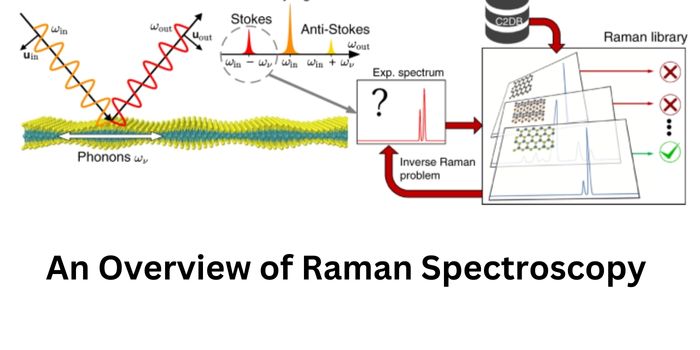
What is The Raman Effect?
Raman spectrometers study changes in monochromatic laser light when interacting with a sample of interest called an analyte. When an event is detected, energy from the incident laser photons is transferred to the analyte. The emitted photons will then experience a shift in frequency. Known as Raman scattering or Raman effect, this inelastic form of scattering can only affect 1 in 10 million photons.
A Raman event that results in a reduction in the energy of an emitted photon is called a “Stokes shift.” On the other hand, a switch to higher energy is called an “anti-Stokes shift.” The former is weaker, especially at greater shifts, because they are dependent on molecules with higher vibrational energy states. The latter, on the other hand, is the most common type of energy used in Raman applications.
Raman spectroscopy is often used to complement infrared absorption spectroscopy. However, these two procedures do not observe similar vibrational information of a molecule. While Raman spectroscopy is based on an inelastic scattering process, infrared spectroscopy is based on the absorption process.
How Is Raman Spectroscopy Useful?
Since it was discovered by C.V. Raman in the 1920s, Raman spectrometers were utilized in fields that require nondestructive chemical and imaging analysis. These spectrometers are used in various industries, from pharmaceutical manufacturing to mineralogy and geology. There are different types of Raman spectrometers ranging from benchtop to handheld.
In the pharmaceutical industry, Raman spectroscopy is used in research and drug development as well as in manufacturing. These spectrometers are used in the qualitative and quantitative investigations of the chemical of interest. In addition, Raman Spectroscopy is used in imaging the analyte as an alternate procedure to various techniques such as HPLC, optical imaging, and NMR, to name just a few.
Aside from the pharmaceutical industry, Raman spectrometers are also used in other fields, which include the following:
- Chemistry – used in monitoring the structure, purity, and reaction of chemicals
- Geology – identification and distribution of minerals, phase transitions, and fluid inclusions
- Life sciences – diagnosis of diseases, drug interactions, and single cells and tissue
- Art and archaeology – characterization of pigments, gemstones, and ceramics
- Semiconductors – intrinsic stress.strain microscope, alloy composition purity
- Pharmaceuticals – uniformity of content and distribution of components
In bacterial infections, Raman spectrometers are helpful in the immediate identification of bacterial pathogens and profiling of antibiotic resistance, which could hasten the precise treatment strategy of infectious diseases.
Raman spectroscopy has also been used to rapidly identify the chemical composition and structure of a sample whether solid, gas, liquid, slurry, gel, or powder.
Types of Raman Spectroscopy
The development of various types of Raman spectrometers has led to improved usability of the instrument. Let us take a look at the different types of Raman spectrometers that have emerged over the years:
- Transmission Raman Spectroscopy (TRS) allows in-depth surveying of analytes, such as tablets and capsules
- Fourier-transform Raman Spectroscopy (FT-Raman) – designed to acquire fluorescence-free and more intense spectra. This type of spectroscopy yields better wavelength accuracy and spectral resolution
- Spatially offset Raman spectroscopy (SORS) utilizes random scattering of light on a turbid media to allow sub-surface surveying. This type of spectroscopy can be beneficial in identifying chemicals through transparent and non-transparent barriers
- Surface-enhanced Raman Spectroscopy (SERS) is an effective technique because it enhances both the laser and Raman signal derived from laser excitation
- Resonance Raman Spectroscopy (RR Spectroscopy), which simplifies the spectrum of particular groups, is used to improve the detection of molecules at small concentrations or to detect the presence of large proteins.
- Coherent Raman Scatterings use multiple laser frequencies to excite the analyte and boost subsequent Raman signals
- Raman spectroscopy that is enhanced by the use of a metal-coated, atomically sharpened tip is known as tip-enhanced Raman spectroscopy (TERS). Raman microscope performance has been improved by spectrometer developers using SERS.
TERS are renowned for precisely regulating the location of the SERS phenomenon and overcoming the spatial resolution constraint of more traditional optical and Raman microscopes.